This study was approved by the Lu'an Traditional Chinese Medicine Hospital Ethics Committee (LASZYY-LL-2023013) and conducted in accordance with the Declaration of Helsinki. Due to the retrospective nature, the requirement for informed consent was waived by the Ethics Committee of Lu'an Traditional Chinese Medicine Hospital.
Study population
A retrospective review of all patients diagnosed with ICH at Lu'an Traditional Chinese Medicine Hospital from February 2017 to January 2023 was conducted. ICH was determined using non-contrast CT images showing parenchymal hemorrhage. Participation criteria: (1) Age > 18 years. (2) Have a history of hypertension. (3) ICH on non-contrast CT scan. (4) Received craniotomy within 72 hours of onset of symptoms. (5) No history of head trauma. (6) Complete clinical data. Exclusion criteria: (1) History of neurological disease such as brain tumor, cerebral infarction, cerebral aneurysm, or other neurological disease. (2) his ICH due to factors other than hypertension; (3) Images of low quality that cannot be evaluated. (4) Suffering from hemophilia, leukemia, or other blood disease. (5) Being treated with anticoagulants or antiplatelet drugs. (6) Associated with primary and secondary coagulopathy. According to the inclusion and exclusion criteria, 609 patients were enrolled in our study. 426 patients from February 2017 to August 2021 participated in the training cohort, and 183 patients from September 2021 to January 2023 were assigned to the external validation cohort (Figure 1) . HICH is the rupture and hemorrhage of blood vessels in the brain parenchyma caused by persistently elevated or wildly fluctuating blood pressure.

Flowchart of HICH patient selection and cohort distribution for predictive model development and validation. ICH intracerebral hemorrhage, hitch hypertensive intracerebral hemorrhage, CT computed tomography, sharp Shapley's additional explanation.
The clinical characteristics and non-contrast CT imaging findings of each patient were meticulously documented. The diagnosis of ICH was confirmed by a baseline non-contrast CT scan within 12 hours of symptom onset. Craniotomy was performed within 72 hours of symptom onset. After surgery, at least two follow-up CT scans of her were performed during her subsequent 3 days.According to the criteria of previously published studies15,27We defined postoperative rebleeding as an increase in hematoma volume of 33% or more compared to the previous postoperative CT scan (ICH volume had significantly decreased after surgery) or within the focal region on follow-up CT. was characterized as either a dense recurrence of Scan after complete surgical removal.
Image acquisition
Baseline noncontrast CT scans (General Electric Medical Systems, Milwaukee, WI, USA) were performed according to standard clinical parameters, utilizing 5-mm-thick axial sections. Images were captured and archived for subsequent analysis. Two experienced neuroimaging experts independently reviewed all images.
Image segmentation and feature extraction
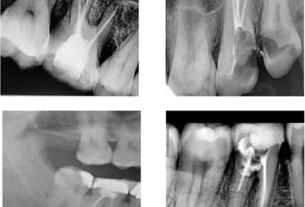
Baseline non-contrast CT images obtained from the Picture Archive and Communication System (PACS) were saved as DICOM files. An experienced radiologist (R1) delineated the region of interest (ROI) using the semi-automated segmentation software 3D Slicer (version 5.0.2) without accessing clinical data (Figure 2) . Her second radiologist (R2) with over 10 years of experience independently confirmed these summaries using the same methodology as R1, again without looking at the clinical data. The consistency of her ROI delineation between observers was assessed by the intraclass correlation coefficient (ICC), with values ≥0.80 indicating high reproducibility. Radioactive signatures from each ROI were extracted using PyRadiomics (version 3.7). Overall, six image types and her six feature classes were obtained.

Non-contrast CT images were semi-automated segmented. CT Computed tomography.
Data preprocessing
Before developing the predictive model, an important data preprocessing phase was conducted to ensure the unbiasedness of the process. At this stage, all data, including both radiomic features and clinical information, were standardized. Standardization depends on the type of data. Continuous variables were normalized using Z-scores to achieve a mean of 0 and a standard deviation of 1, whereas categorical variables were converted to binary form and labeled as “0” or “1” .
Radiomics function selection
We performed three feature selection steps to reduce overfitting. Initially, an ICC greater than 0.8 was considered to indicate satisfactory agreement. continue, t-A test was applied to each feature to distinguish patients with and without postoperative rebleeding. Finally, using Least Absolute Shrinkage Selection Operator (LASSO) logistic regression combined with 10-fold cross-validation, features associated with postoperative rebleeding with non-zero coefficients were selected from those exhibiting a P value less than 0.05. I did. t-test.
ML model derivation and internal validation
To assess the risk of postoperative rebleeding, we utilized four established ML classifiers: Logit, RF, support vector machine (SVM), and XGBoost. We used clinical data, radiomics features, and their combination to create well-defined predictive models. During training, we applied 5-fold cross-validation with three iterations to enhance data utilization and divided the training set into internal training and testing subsets for sequential evaluation. For the RF model, we chose a 500-tree configuration and determined the number of features for node splitting based on the square root of the total number of features. SVM used a radial basis function (RBF) kernel, which is adept at handling nonlinear data, and fine-tuned hyperparameters through grid search. This also includes overall cost parameters. [0.1, 1, 10] and the gamma parameter of the RBF kernel. [0.001, 0.01, 0.1]. XGBoost parameters, such as a learning rate of 0.02, a maximum tree depth of 4, and a 600 tree ensemble, are optimized through a grid search to ensure a delicate balance between prediction complexity and accuracy and streamline the model development process.
After deriving each model, we conducted a rigorous internal validation process to assess its identification, calibration, and clinical applicability. The selection of the optimal predictive model was based on its excellent discriminatory power, along with robust calibration and clinical utility.
Interpretability and external validation of ML models
After identifying the best predictive model, we shifted our focus to understanding the contribution of each variable to prediction. We incorporated the SHAP (Shapley Additive Preparation) technique to gain deeper insight into the importance of features while highlighting the most influential variables. Features were ranked by SHAP value in descending order of influence to accurately identify key predictors within the patient cohort. To ensure the robustness of the model, we conducted external validation. This thorough evaluation confirmed the discriminatory power, calibration, and clinical relevance of the models and provided a comprehensive perspective on the predictive strength of these models.
statistical analysis
Statistical evaluations were performed using R statistical software (version 4.2.1) and Python programming software (version 3.7.1). Continuous variables that showed skewed distributions were expressed as medians. [interquartile range (IQR)] Evaluation using the Mann-Whitney method U-test. Categorical data are presented as numbers (percentages) and analyzed using χ.2 test. Model performance evaluation includes receiver operating characteristic (ROC) curve analysis focusing on area under the curve (AUC) and metrics such as precision, recall, and F1 score for a thorough evaluation of discrimination ability. included. His Delong's test was used to compare AUC. Model fit was assessed using calibration curve analysis and Brier scores for probability prediction accuracy. To assess the clinical utility of the model, we used decision curve analysis (DCA) to calculate the net benefit across different threshold probabilities.
ethical approval
This study was approved by the Ethics Committee of Lu'An Hospital of Traditional Chinese Medicine (number LASZYY-LL-2023013).
informed consent
Written informed consent was waived by the Ethics Committee of Lu'an Traditional Chinese Medicine Hospital.