image:
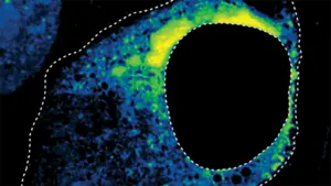
Microscopic image of cells infected with respiratory syncytial virus treated with antiviral drugs. This drug causes a change in the shape of the viral condensate (shown in yellow), thereby inhibiting viral replication. The large black circular structure is the nucleus of the cell. Image provided by: researcher
view more
Credit: Cliff Brangwyn, Princeton University
Researchers at Princeton University have introduced a tool that leverages AI to understand how drugs affect the dynamics of important structures within cells, mapping the shape of these structures to functional outcomes, and can shed light on important markers of health.
To do this, a team led by Cliff Brangwyn examined changes in the shape of biomolecular condensates, tiny droplets inside living cells that drive transcription and other gene regulation processes and are associated with diseases such as Alzheimer’s disease, ALS, and cancer.
“A central question in biology is how to obtain emergent structures from the interactions of individual molecules,” said Brangwyn, the June K. Wu ’92 Professor of Chemical and Biological Engineering and the study’s principal investigator. “The key innovation here was to develop a way to learn from images and classify the patterns that emerged.”
The study, published June 4 in the journal Cell, focused on condensates called nucleoli, which are responsible for assembling tiny machines that build proteins. The research team used an advanced microscope to image changes in the shape of the nucleolus in hundreds of human cells under a variety of drug-controlled conditions. They then fed these images, which are difficult for even highly trained scientists to interpret, through a machine learning tool they had built for this purpose. This tool was able to classify images into four basic categories based on the shape of the nucleolus. Three were categories the researchers had expected, and the fourth was unexpected and was later determined by the team to be an entirely new category.
These cap and necklace shapes have been associated with a variety of cellular stress responses, making them useful markers for testing the efficacy of drugs and gene therapies. For example, the cap shape could result from a process known to disrupt the process that creates the RNA needed to assemble tiny protein-making machines. The necklace shape may arise from a class of drugs that interfere with different RNA-related processes.
After initial training, the team conducted a drug panel study to see how each drug affected nucleolus formation. They used a neural network to visually measure changes in condensate development and found that different concentrations of the drug caused different degrees of change in both the cap and the necklace.
The neural network found that two known cancer drugs cause capping, a phenomenon not previously reported for these drugs, said postdoctoral researcher Anita Donric, lead author of the paper. This suggests that these drugs may affect nucleolar function in previously unrecognized ways.
For the third drug, called topotecan, the network discovered an entirely new nucleolar shape, which the researchers named a flower.
Although topotecan was known to inhibit enzymes used in DNA replication, Donric showed that deletion of this enzyme, TOP1, induced flower shape, revealing the enzyme’s role in maintaining nucleolar organization by regulating RNA processing.
“No one has ever seen this flower form before,” said Brangwyn, who is also director of the Ormen-Darling Institute for Bioengineering. “The network flagged it as not fitting neatly into the other three categories.”
These findings point the way toward robust systems for monitoring and evaluating cellular responses to drugs at the single-cell level.
The researchers also tested the neural network on other condensates associated with RNA processing and observed similar dose-response results for drugs specific to nuclear speckles (a hub of messenger RNA activity) and respiratory syncytial virus condensates.
This discovery highlights the need to unravel molecular-level mysteries about fundamental factors such as size and shape that are often overlooked by human analysis. “Other important features may be missing,” Donlic says. “Something that could indicate the existence of new biology.”
The paper “Deep learning of functional perturbations from condensed morphology” was published in the journal Cell on June 4, 2026. In addition to Brangwynne, Donlic, and Comi, authors include Sofia Quinodoz, Nima Jaberi-Lashkari, Krist Antunes Fernandez, Lifei Jiang, Lennard Wiesner, and Ai Ing Lim of Princeton University. Support for this project was provided in part by the Howard Hughes Medical Institute, the Princeton Composite Materials Center (NSF MRSEC DMR-2011750), the St. Jude Membraneless Organelle Collaboration, the Chan Zuckerberg Initiative Exploratory Cell Network, and the Princeton Institute for Artificial Intelligence.
Research method
experimental research
Research theme
cell
Article title
Deep learning of functional perturbations from condensate morphology
Article publication date
June 4, 2026
Disclaimer: AAAS and EurekAlert! We are not responsible for the accuracy of news releases posted on EurekAlert! Use of Information by Contributing Institutions or via the EurekAlert System.


