Cameras used for patient ID typically do not look like medical tests.
But in a new cancer study, regular mugshots taken months or years apart seemed to capture something deeper. That is, how quickly a person ages biologically while undergoing treatment and how that change is tracked in survival.
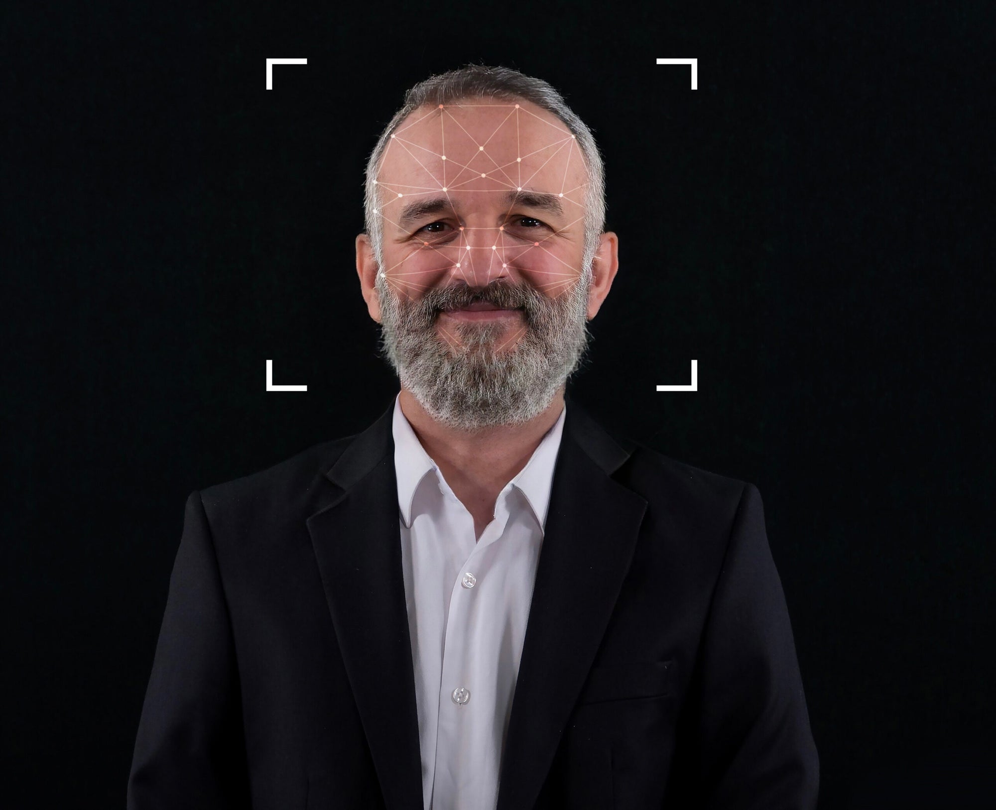
Researchers at Massachusetts General Brigham Research Institute say this changing metric, called facial aging rate (FAR), could offer a new way to read a patient’s health over time without drawing blood, scanning, or biopsies. Writing in the journal Nature Communications, the research team reports that cancer patients whose faces age faster than expected are more likely to die earlier than those whose faces age more slowly or remain constant.
The study builds on previous research behind FaceAge, an artificial intelligence tool that estimates biological age from a single facial photo. The group reported last year that cancer patients often look about five years older than their actual age, and that higher FaceAge estimates were associated with lower survival rates after treatment.
This time, the question wasn’t how old someone looked in an image. It was the speed at which the appearance of age changed.
When the face begins to act like a biomarker
The study examined a series of facial photographs of 2,276 cancer patients age 20 and older who had received at least two courses of radiation therapy at Brigham and Women’s Hospital. These photos are already part of the routine clinical workflow and were taken at the start of each radiotherapy course from 2012 to 2023.
The researchers calculated FAR by comparing two images from each patient (one from an earlier treatment time point and one from a later treatment time point), measuring the change in FaceAge, and dividing it by the time between the photos. Values above 1 suggest accelerated aging. Values less than 1 indicate slow aging.
We also tracked FaceAge Deviation (FAD). This is another measure of how old or young a person looks in a single image compared to their actual age.
Across the group, the median age at first radiotherapy course was 63.4 years, and patient age ranged from 20.1 to 97.0 years. Women made up 50.5% of the cohort and men 49.5%. Most patients were white (85.1%), followed by black patients (5.1%), Asian patients (4.9%), and patients reporting other races (4.9%).
The median time between two photos was 286 days. Median follow-up was 35.7 months.
What was most noticeable was the speed of change. Median FAR results suggested that patients’ facial aging exceeded chronological aging by 40%.
Facial aging is accelerated and life expectancy is shortened.
The research team divided the patients into three groups based on the spacing between the photos. One is short term, from 10 days to 365 days. medium term, 366 to 730 days; Long term from 731 days to 1,460 days.
The timing was important. FAR fluctuates widely over short intervals, but because the denominator is small, even small fluctuations can result in large changes. At longer spans, the values appeared narrower and more stable.
Still, the survival signal was retained in all three groups.
Among patients in the short-interval group, those with high FAR had a median survival of 4.1 months compared to 6.5 months for those with low FAR. In the intermediate group, patients with early facial aging had a median survival of 6.4 months compared to 12.5 months for those with slow facial aging. In the long-term group, the difference was even wider: 15.2 months vs. 36.5 months.
Hazard ratios showed similar results. In univariate analysis, higher FAR was associated with higher risk of death in all three time windows. For example, in the long-interval group, the hazard ratio for a FAR greater than 1 was 1.60. After adjusting for time between photos, gender, race, and cancer diagnosis at second radiation therapy, the association remained significant, with an adjusted hazard ratio of 1.65.
This pattern also held true for patients with metastatic cancer at the time of their second radiation treatment, the highest risk subgroup in this study. The authors said the divergence in survival curves was even more pronounced there.
Not all regular markers behave the same way. Age itself showed no significant association with survival in any time interval group. Ethnicity and diagnosis were differentially associated depending on cohort, and male sex was associated with a lower risk of death only in the long interval group.
Moving measures may be more important than snapshots
One of the most interesting results of this study came from comparing FAR with the one-time FaceAge Deviation measurement.
Patients with both high FAD and high FAR faced the worst survival prospects. However, over time, FAR has come to provide more useful information than FAD alone. In the short-interval group, both measures contributed to mortality risk. At longer intervals, the rate of aging itself appears to have greater weight in prognosis.
This is important because it suggests that the pace of visible biological change may indicate a person’s apparent age more than a single estimate.
The research team confirmed this change in contour plots that map hazard ratios across combinations of FAD and FAR. In short-term intervals, both values were significant. At longer intervals, the contours flatten, indicating that the FAR has become a stronger signal.
Additional analyzes using later FAD values showed similar trends, although later one-time measurements did not distinguish results as clearly as FAR. The authors write that extended analysis showed that FAR outperformed single-time point FAD across all time intervals, with the strongest performance in the long-term group.
“Deducing facial aging rates from multiple daily facial photos allows us to track an individual’s health status in near real time,” said co-senior and corresponding author Raymond Mack, a radiation oncologist at Massachusetts General Brigham Cancer Institute and faculty member in the system’s medical artificial intelligence program. “Our study suggests that measuring FaceAge over time may improve individualized treatment plans, improve patient counseling, and help guide the frequency and intensity of follow-up in oncology.”
What faces recognize
The authors argue that the progression of facial aging may reflect broader biological distortions, including cellular senescence, DNA damage, and reduced tissue repair, all of which are linked to both aging and cancer progression.
However, the face itself does not explain the disease. This means that visible changes may be acting as a proxy for other underlying health changes that unfold.
This group frames FAR as a dynamic biomarker. This may be particularly useful because in medicine we often learn more from repeated measurements than from a single measurement. The article points out similar patterns in other fields, from blood pressure fluctuations in cardiovascular care to PSA velocity in prostate cancer to repeated biomarker tracking in Alzheimer’s disease.
There’s a practical appeal here too. Unlike many biological age measurements, FAR requires no laboratory analysis or specialized equipment beyond clinical photography, which is already used in some cancer settings.
That might make it easier to repeat.
The researchers noted that many patients in the cohort had metastatic disease, 62.9% at the first course of radiation and 78.7% at the second course. In that context, a high FAR may help identify people who require less aggressive and less toxic palliative treatment rather than escalation. It also helps clinicians more carefully balance symptom relief, quality of life, and survival goals.
A second recent FaceAge study, published in The Lancet Digital Health, furthered the broader idea. In more than 24,500 cancer patients aged 60 and older who received radiation therapy, FaceAge revealed that 65% of patients were older than their actual age. Those whose FaceAge estimates were at least 10 years older than their actual age had poorer survival outcomes, and those whose FaceAge estimates were within 5 years had better outcomes.
The limit is not small
Although the results of this study are promising, there are obvious limitations to this study.
The patient cohort was primarily Caucasian, which could limit how generalizable the results are to more diverse populations, the authors note, as facial aging patterns in particular can vary by racial group. Most of the patients are also elderly, and open questions remain as to whether the results look the same in younger people.
Photo quality, lighting, and facial expressions can affect performance. Researchers also lack detailed data on disease progression, treatment details, cachexia, and toxic effects, all of which can impact both appearance and survival. These missing variables may have acted as confounders.
There’s also the issue of timing. Photos were not taken at regular study intervals. Because these were captured at specific radiotherapy time points, the short-term, intermediate-term, and long-term groups may reflect different clinical situations rather than a precise clock-based comparison.
And although the association was significant, the model has not yet been tested in a prospective clinical trial.
Next, there are ethical issues. The article points out privacy concerns, the potential for bias in AI systems that analyze faces, and the need for transparent algorithms and strong data protection before wider clinical use.
“Long-term tracking of FaceAge from a simple photo provides a non-invasive, cost-effective biomarker that has the potential to inform an individual’s health status,” said co-author Hugo Aerts, director of the Medical Artificial Intelligence Program at Massachusetts General Brigham. “We hope that with continued research, we can learn how FaceAge can provide prognostic information to patients with other chronic diseases and to healthy people.”
Practical implications of the research
At this time, FAR is not an independent decision-making tool, and this study does not show that facial aging changes alter patient outcomes. The study suggests that repeating face-based age estimation could add a new layer to cancer treatment, especially as doctors seek to understand how patients are holding up over time.
In oncology, this could mean better risk stratification, better follow-up, and clearer conversations about treatment intensity. In the long term, the researchers believe the same approach could be tested in chronically ill and healthier populations, with repeated non-invasive tests potentially detecting changes in health before symptoms become apparent.
The team has also launched an Institutional Review Board-approved web portal where the public can submit photos to be evaluated by FaceAge and contribute to research. Whether this technology becomes part of routine care will depend on something less flashy than the algorithm itself: careful validation, unbiased performance across populations, and proof that the information improves real-world clinical decision-making.


