
FRANCOIS NASCIMBENI/AFP via Getty Images
For a surgeon removing a tumor from the pituitary gland, the margin between success and catastrophe is literally about 1 millimeter (0.04 inches) — the distance separating the optic chiasm, the major blood vessels, and the nerves that control vision from a tumor that has no business being there. In May 2026, a team at London’s National Hospital for Neurology and Neurosurgery crossed a line that surgical AI had been approaching for years: they deployed a real-time computer vision system in a live human operation, using it to color-code the anatomy around a patient’s pituitary gland as surgeons removed his tumor — and the patient woke up seeing clearly for the first time in over a year.
The Patient and the Tumor
Rhys Hibbert, 48, from Bedfordshire, England, discovered he had a brain tumor by accident. He had collapsed during a walk in December 2024 and suffered a seizure — the first of his life. Hospital tests revealed an 11-millimeter pituitary adenoma: a non-cancerous tumor on the pituitary gland, a marble-sized organ at the base of the skull that regulates nearly every major hormone in the body.
Pituitary adenomas of this size — technically classified as macroadenomas — are common in the clinical sense, affecting approximately 1 in 1,000 people at levels requiring treatment. What makes them surgically treacherous is pure anatomy. The pituitary sits in a bony depression called the sella turcica, packed in alongside the optic chiasm (the nerve-crossing point where damage causes bilateral peripheral vision loss), the cavernous sinuses, and the internal carotid arteries. A millimeter of error in any direction can mean blindness, stroke, or death.
Hibbert’s tumor was initially managed without surgery, but his symptoms worsened progressively over more than a year — severe hormone imbalance, deteriorating vision, and eventually an inability to see the lower field of his visual range. He began using walking sticks after repeatedly tripping. When surgery became unavoidable, he was asked whether he would volunteer to be the first patient to have his procedure performed with a live AI surgical assistant. He agreed, according to UCLH.
How the System Works
The AI was built at UCL’s Hawkes Institute, a multidisciplinary research group at the intersection of UCL’s engineering and health activities, and is designed to run on NVIDIA’s Clara IGX platform — purpose-built hardware for real-time AI inference in medical device environments, designed to meet IEC 62304 software lifecycle requirements and deliver deterministic, low-latency processing at the surgical edge.
The core of the system is a convolutional neural network (CNN) trained on a large collection of annotated endoscopic pituitary surgery videos from previous operations. Neurosurgeons watched those videos and labeled each frame — marking where the sella is, where critical vessels run, where the tumor boundary sits — using polygon annotation tools to create the ground truth the model learned from. The trained model then performs semantic segmentation on live video: frame by frame, it classifies each pixel in the camera feed as belonging to a specific anatomical structure, and overlays that classification as color-coded labels on a secondary monitor beside the primary surgical display.
This is categorically different from the image-guided navigation systems that neurosurgeons have used for years. Those systems register pre-operative MRI scans to the patient’s physical anatomy and give surgeons a static map. The problem is brain shift: as surgery proceeds, cerebrospinal fluid drains, tumor is removed, and the brain physically moves — by several millimeters in some cases, enough to invalidate a static map precisely when the surgeon needs guidance most. The UCL system sees what the surgeon sees, updated in real time, unaffected by any tissue movement that has happened since the patient entered the operating room.
Dr. Sophia Bano, Associate Professor in Robotics and Artificial Intelligence at UCL Computer Science and the technical lead for the project, described the system’s reach: according to the UCL announcement, “By learning from hundreds of surgical videos, it has been exposed to a breadth of surgical examples that would take a surgeon many years to encounter. It is designed to help recognize critical anatomy, surgical instruments and tissue interactions in real time, supporting the surgeon during highly delicate procedures.”
What the Published Research Showed
The UCL team did not deploy this system speculatively. The work behind it spans years of published research. An earlier version of the anatomy recognition model was evaluated in a study published in npj Digital Medicine in November 2024, testing its effect on clinicians’ ability to correctly identify the sella — the key surgical landmark — in still endoscopic frames.
The results were measurable and pointed to something important. Overall, AI assistance improved anatomy recognition accuracy from a DICE score of 70.7% to 77.5% — a 6.7 percentage point improvement that was statistically significant. But the improvement was not evenly distributed across experience levels. Medical students — the least experienced group — improved from 66.2% to 78.9%, a gain of 12.8 percentage points. Senior surgeons still benefited, but less. The system’s value is inversely proportional to the operator’s existing expertise.
This pattern has significant implications beyond any single operation. Pituitary surgery is a low-volume, high-difficulty specialty: it is concentrated at major tertiary centers precisely because the learning curve is steep and the anatomy unforgiving. A surgeon in a hospital that performs dozens of these procedures a year, rather than hundreds, faces an experience gap that pre-operative MRI and intuition cannot fully close. A system trained on the full spectrum of cases from many operators and made available at the point of surgery could, in principle, narrow that gap. The largest benefit of this AI, the data suggest, is not for the world’s most experienced pituitary surgeons — it is for the ones who could use the most support, and by extension, for the patients who cannot travel to the handful of institutions where those experts practice.
What Happened in the Operating Room
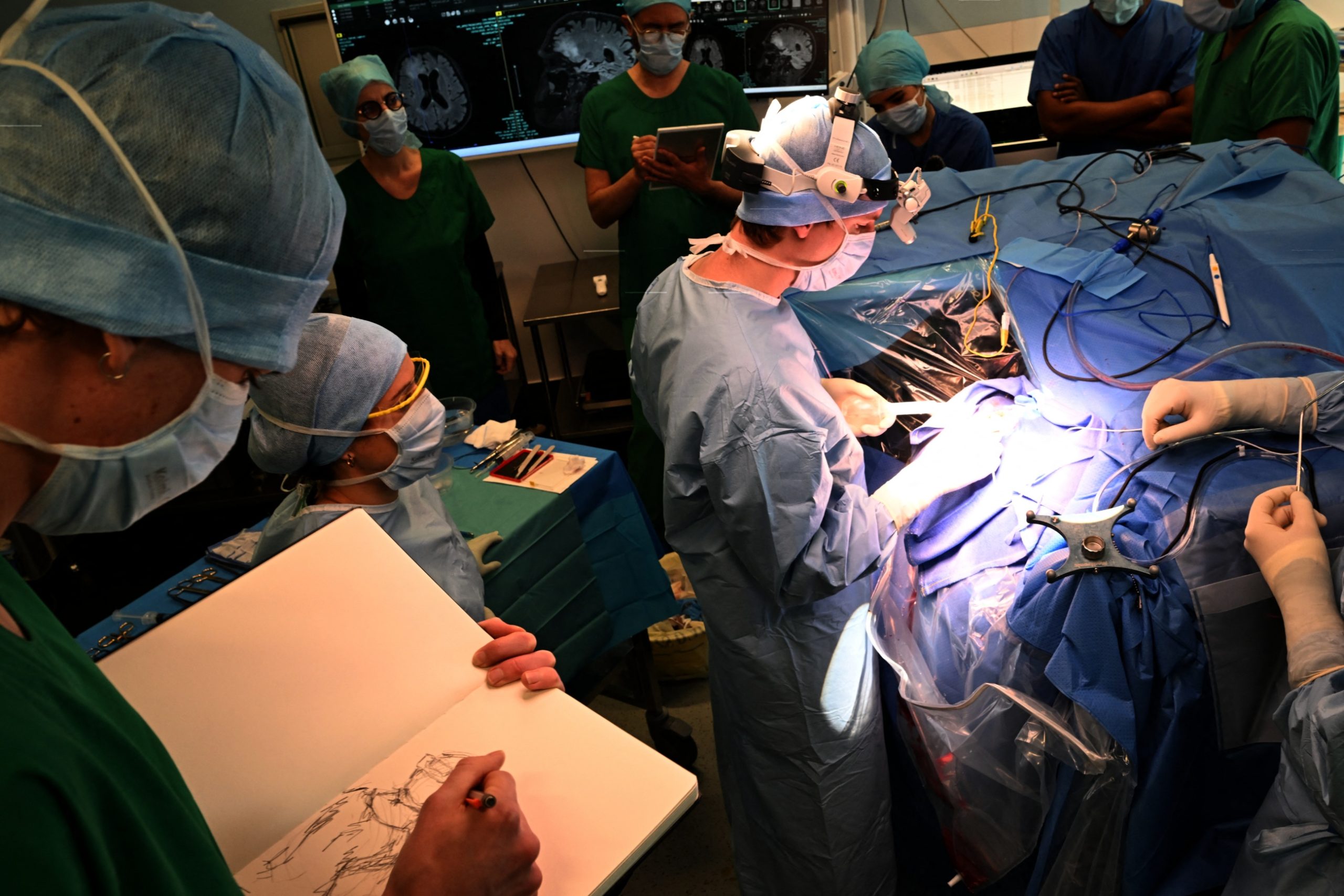
Professor Hani Marcus, of the UCL Queen Square Institute of Neurology and a consultant neurosurgeon at the NHNN, led the operation alongside Mr. Danyal Khan, a UCL PhD candidate and UCL/UCLH neurosurgical resident who has led the research program driving this technology toward clinical use. During surgery, the AI analyzed the live feed from the endoscope — the thin tube carrying a camera through the patient’s nostril and sphenoid sinus to the base of the skull — and displayed its anatomy labels on a secondary monitor. It did not control any instrument. It did not make any decision. It functioned as what Professor Marcus called an expert second pair of eyes that never tires, never loses concentration, and has seen more operations than any human surgeon alive.
When Hibbert woke from anesthesia, his vision had dramatically improved. Within a week, he was walking independently without glasses or sticks. “It feels like I’ve got a 360-degree panoramic view of everything around me,” he said — something he had not experienced for more than a year. He is now back at work as a customer services manager and continues volunteering as a community ambulance first responder.
Why a Secondary Monitor Matters
The current system presents its guidance on a screen beside the main surgical feed rather than directly overlaid onto it. This means surgeons must divide their visual attention between two displays during the most delicate moments of the operation — a real engineering constraint that the research team has already identified as a target for future refinement. The next generation of the system is also expected to track surgical instruments and monitor tissue-instrument interactions in real time, providing a more dynamic picture of the surgical field beyond static anatomy labeling.
Independent research on intraoperative AI systems has identified consistent challenges across this field: performance degrades when the surgical field is obscured by blood, smoke, or instrument crowding; class imbalance in training datasets means rare anatomical variants may be underrepresented; and systems trained at one surgical center do not always generalize reliably when deployed at another.
The UCL system was used here as a research tool under a clinical trial, funded by NIHR and Google and supported by the NIHR Biomedical Research Centre at UCLH, the Royal College of Surgeons, the Engineering and Physical Sciences Research Council, and Wellcome. It has not been cleared for routine clinical use outside the trial, and the research team will need to expand the trial and validate performance across a broader patient cohort before any path toward standard deployment opens.
Does AI Help Better Surgeons or Surgeons Who Need It Most?
A surgeon survey published in Surgical Endoscopy in June 2026 found that 86.8% of surgeons believed intraoperative AI could positively impact surgical performance, and that anatomy recognition received the highest usefulness scores of any AI function assessed — 4.57 out of 5. Despite this, the same survey found that adoption in routine practice remains limited, with a persistent gap between positive perceptions and clinical implementation.
The UCL anatomy recognition data points to where that gap could matter most. If the technology’s benefit is largest for less experienced operators, its widest impact may not be in the world’s leading neurosurgical centers — which already have the volume and expertise to navigate the sella safely. It may instead be in the next tier of institutions: hospitals that perform pituitary surgery at moderate volume, serve regional patient populations who cannot easily travel, and whose surgeons could benefit from the kind of pattern-recognition breadth that only comes from having seen hundreds of operations spanning many operators and anatomical variations. That is the scale of surgical exposure the UCL system encodes.
UK’s Oldest Neurosurgical Hospital Performs Its Latest First
It was not lost on Hibbert that the institution where his procedure took place carries its own history. The National Hospital for Neurology and Neurosurgery was founded in 1859 as the world’s first dedicated neurological hospital — originally called the National Hospital for the Paralysed and Epileptic — by Johanna Chandler and her siblings, who raised funds to care for those suffering from paralysis and epilepsy at a time when no such institution existed.
Today it is the United Kingdom’s largest dedicated neurological hospital, treating more than 6,000 inpatients and 180,000 outpatients a year. “The NHNN was founded in 1859… and was the world’s first dedicated neurosurgical hospital,” Hibbert said. “So to me, it seems very fitting that the same hospital should also be the world’s first to carry out an AI-assisted neurosurgery.”
James Frith, the UK’s Health Innovation Minister at the Department of Health and Social Care, endorsed the achievement: “This is an example of AI at its best: patients getting care previously deemed unimaginable thanks to the latest groundbreaking technology. AI needs proper safeguards and we will always ensure that safety is taken seriously. But we will also ensure we benefit from the opportunities it brings to deliver faster and more effective care.” Frith previously served as a minister in the Department for Science, Innovation and Technology, where he attended the launch of the Cambridge Zenith AI Supercomputer — the UK’s largest AI-for-science platform — before moving to his current health brief in July 2026.
Frequently Asked Questions
How does the AI surgical system at UCL actually know where not to cut?
The system uses a convolutional neural network — a type of deep learning architecture — trained on hundreds of annotated endoscopic pituitary surgery videos. During those surgeries, expert neurosurgeons labeled each video frame to identify the sella (the bony landmark containing the pituitary gland), surrounding blood vessels, and nerves controlling vision. The trained model learned to recognize these structures by their appearance in live video. During surgery, it processes the endoscopic camera feed in real time, classifies each pixel as belonging to a specific anatomical structure, and displays color-coded labels on a secondary monitor beside the surgeon’s primary view. It does not control any instrument or make any decision — it provides a visual overlay the surgeon can use to orient themselves in one of neurosurgery’s most congested anatomical environments. Full technical details are available through the UCL Hawkes Institute.
Why does AI help less experienced surgeons more than expert neurosurgeons?
In clinical tests of the UCL anatomy recognition system, experienced surgeons improved their anatomical accuracy by a moderate amount with AI assistance — but the biggest gains went to less experienced clinicians, including medical students, who improved by 12.8 percentage points compared to 6.7 percentage points overall. Expert surgeons have already internalized the pattern recognition the AI encodes; less experienced surgeons have not yet accumulated enough operative experience to match it. This is a critical finding for the technology’s long-term impact: if a system trained on hundreds of cases can partially compensate for limited individual experience, it could one day help reduce the outcomes gap between high-volume specialist centers and the regional hospitals where most patients actually receive care. The full data are published in Khan et al., npj Digital Medicine.
Can this AI be used in other types of surgery, not just pituitary procedures?
The current UCL system was trained specifically on endoscopic pituitary surgery — a narrow procedure type with consistent anatomy and a clearly defined surgical corridor. Generalizing it to other procedures would require new annotated training datasets covering those surgeries, as well as validation studies demonstrating safe performance in each new context. The research team and the broader surgical AI community have identified generalization — the ability to perform reliably across different surgical centers, equipment setups, and procedure types — as one of the major unsolved challenges in this field. That said, the underlying framework (CNN-based real-time semantic segmentation on live surgical video, running on NVIDIA’s Clara IGX platform) is designed to be adaptable, and several companies and research groups are pursuing similar architectures for laparoscopic, robotic, and other endoscopic procedures.
What safeguards ensure the AI cannot cause harm during surgery?
The system is classified as a decision-support tool, not an autonomous agent — it displays guidance information on a secondary monitor, and the surgeon retains full control of every instrument and every decision throughout the operation. The NVIDIA Clara IGX platform on which it runs is purpose-built for safety-critical medical device environments, designed to meet deterministic real-time requirements and long-term software lifecycle standards (IEC 62304). The procedure itself took place under an NIHR-funded clinical trial with appropriate research ethics oversight. Routine clinical deployment would require regulatory clearance from the UK’s Medicines and Healthcare products Regulatory Agency — a process that involves formal validation of safety and performance across a defined patient population. The system has not yet reached that stage; the May 2026 procedure represents the first step in the IDEAL surgical innovation framework’s first-in-human stage.


