
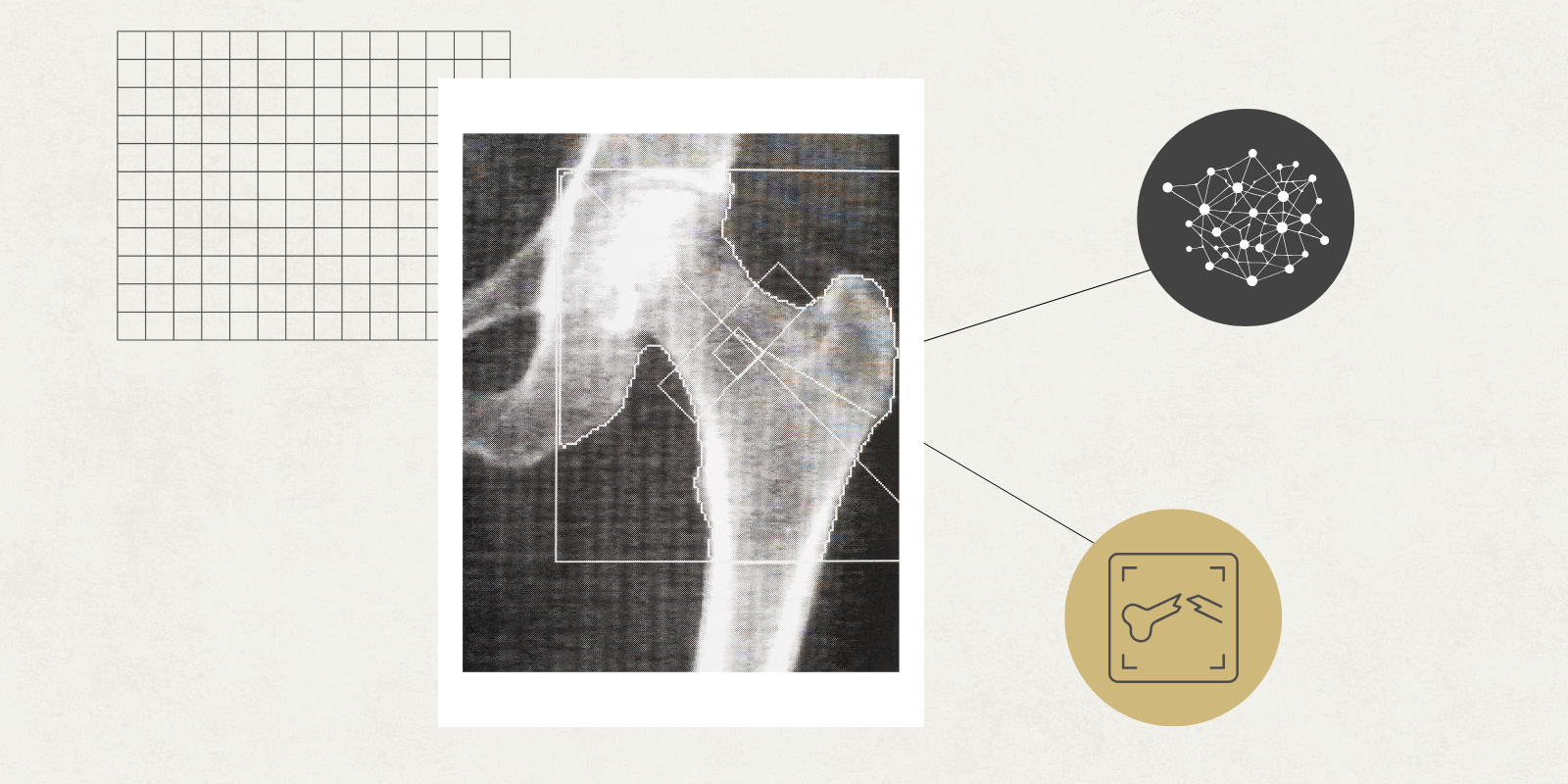
Perhaps in the not-too-distant future, artificial intelligence and its subfield machine learning (ML) tools and models could help orthopedic surgeons predict when and what types of fractures their patients will encounter.
“Currently, these types of tools are not widely used in clinical practice, but they are used in research,” says Dr. Michael A. David, lecturer in orthopedic surgery at the University of Colorado Anschutz School of Medicine. “What’s appealing is that these tools can take large amounts of data and make sense of it efficiently with minimal human input.”
The use of machine learning – a broad field of computing for “teaching” computers to automate tasks and discover unexplored data patterns – is expected to accelerate these efforts, and David’s research is helping to advance them.
His research integrates ML with spatial histopathology, transcriptomics, and medical imaging to study and diagnose post-traumatic and idiopathic joint conditions such as contractures, osteoarthritis, osteoporosis, and tendon ruptures. This job often requires a deep familiarity with the medical knowledge required in orthopedics and the technical expertise that enables the construction and operation of ML tools.
In a new review article published in bone reportDavid explains, “Machine learning has the potential to enable agnostic, automated analysis that accelerates discovery and improves clinical bone diagnosis and treatment monitoring.”
Utilizing bone image data using machine learning
One useful application of ML is the ability to perform segmentation (dividing digital bone images into distinct bone types much faster than humans can). Bone imaging provides important data for orthopedic surgery research, which is a major benefit since large amounts of data are required to be effective.
“Segmenting bone data is the most arduous and time-consuming task,” says David, who did the work himself as a graduate student. “This is one area where ML excels in accelerating the pace of discovery: automating segmentation and extracting quantitative data from images.”
However, there are some caveats. Not all bones and images contain the same information, nor are they relevant to all research efforts. ML still requires human expertise and analysis to be effective.
David’s review paper focuses on how researchers are using ML in micro-computed tomography (microCT) and high-resolution peripheral quantitative computed tomography (HRpQCT), two common imaging techniques for assessing bone structure.
In these areas, ML has been used “primarily to improve image resolution, automate bone segmentation, reduce the number of experimental variables, cluster data, and combine these with prediction and classification of bone health and fracture risk,” says David.
Although these technological advances are impressive and meaningful, it is still very unlikely that ML will replace radiologists in orthopedics. Instead, David says these methods will come in the form of clinical tools that help healthcare providers make the best decisions for their patients.
Using data to cluster patient groups can also be beneficial to patients, as they can learn more about subtypes and stages of diseases such as osteoporosis.
“ML allows us to combine different types of data, such as bone images, genetics, and even sleep cycle information, to see if subgroups form. I think there is a lot to be gained from this field in terms of progress,” says David.
Preparing the next wave of scientists
The use of ML is likely to become more prominent in the coming decades, but to be useful, researchers need a deep understanding of ML.
“Especially with the rise of large language ML models such as Deepseek, ChatGPT, Claude, and Gemini, important information is now available in the public domain, but the sheer breadth of tools, models, metrics, and interpretability measures creates a steep learning curve,” says David. “The lengthy and difficult learning process can be a significant barrier to entry for bone researchers looking to explore and implement ML approaches.”
To alleviate that barrier, David wrote a review paper and in the process created reproducible code for basic ML coding. He also developed SciNetX, a scientometric and bibliometric analysis pipeline and visualization software.
“SciNetX was born out of the fact that when completing a review study, you need to come up with a structure and show how all the research papers you looked at fit together,” he says.
You enter different types of information, such as paper subjects, keywords, and authors, and the software creates a visual network that shows how different topics and researchers are related.
“This is basically a map of the site,” says David. “As a new researcher in this field, I was able to look at this paper and see where this research started, who is doing it, and who they are collaborating with.”
David developed this software to work in orthopedics, but it can be applied almost anywhere, from a variety of medical research efforts to those completely outside of medical and scientific research. He is currently working with CU Innovations to license the program and make it available to others.
Ultimately, David envisions a symbiosis of human-computer interaction, and his goal is to incorporate ML into digital bone imaging, lowering the barriers for those who want to uncover vast networks of scientific knowledge.
“The whole idea is to invite more people into this world,” says David. “As we were developing SciNetX, we realized how powerful it is. We want people entering this field of research to be ready to learn, and this will help us achieve that.”

