
Long before artificial intelligence began promising to improve medical diagnostics, a little gray bird was already doing pretty well.
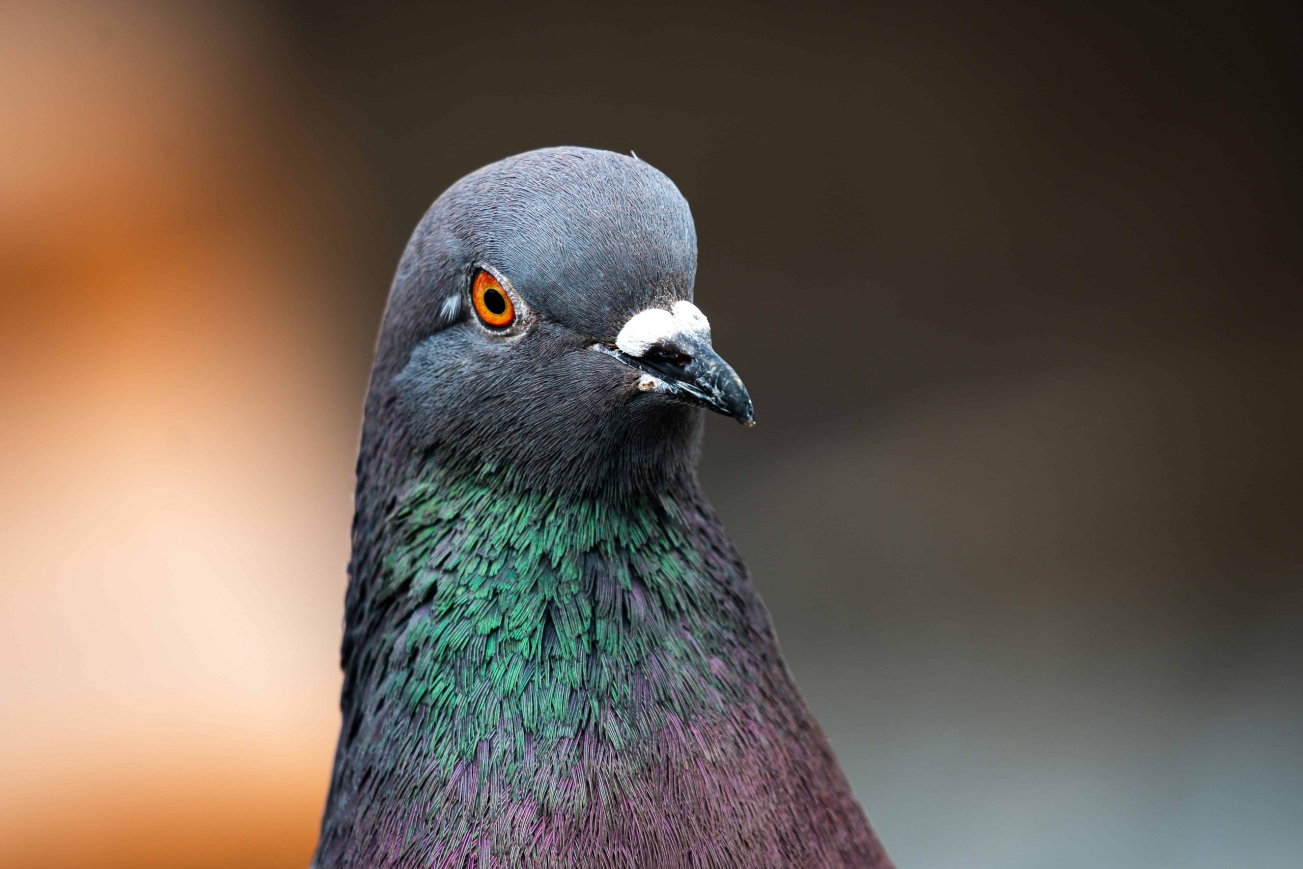
In 2015, pigeons (Columba Libya) They learned to do things that almost sounded stupid. They learned to look at images of breast tissue and tell the difference between benign and malignant samples. With training, its accuracy has become comparable to that of experts. In fact, after about two weeks of training, individual pigeons reached about 85% accuracy in distinguishing between cancerous and non-cancerous breast tissue.
But it’s even better. When they put their brains together, they achieved a whopping 99% accuracy on one diagnostic task.
A bird brain made for seeing
Pigeon brains are small, perhaps no larger than the tip of a human’s index finger. But just because it’s small doesn’t mean it’s simple. And it turns out they’re very good at recognizing patterns. Pigeons have been shown to recognize human faces and emotional expressions, letters of the alphabet, distorted pharmaceutical capsules, and paintings by Monet and Picasso.
In a 2015 study led by Professor Richard Levenson of the University of California, Davis, and Professor Edward Wasserman of the University of Iowa, pigeons were trained to examine digital images of breast biopsy slides. Each bird learned to face a screen and peck at a button representing benign tissue and another button representing malignant tissue. Those who answered correctly were given food pellets.
First of all, it turns out that their memory is amazing. The pigeons were able to recall more than 1,800 images, and in some cases very similar images. But the point was not memorization, but recognizing new images that had never been seen before.
The birds were looking for visual hallmarks of malignant tumors: darker, denser, abnormal-looking cell patterns. On selected breast biopsy and mammography tasks, the trained pigeons approached the performance of human experts. But when the researchers combined the decisions of the four birds (which, interestingly, they called “flock sourcing”), they pushed performance to 99% accuracy.

Admittedly, these were controlled images that failed at the more difficult task of determining suspicious mammographic masses. But it’s a surprising result.
More than 10 years have passed since this study was published, but no pigeon team exists to screen mammograms. But this begs the question. Why are they so good?
have received prior training
First of all, this skill makes evolutionary sense. Pigeons survive by quickly scanning cluttered scenes, including gravel, dirt, leaves, shadows, food, and predators. There are a lot of small visual differences in that world that are important. Seeds hidden among pebbles are viable targets, and small movements overhead can signal danger.
The birds viewed images of breast tissue at different magnifications, much like a pathologist would examine a slide under a microscope. They learned how to classify full-color images, and some were also trained on modified images with reduced color cues. They were also tested for different levels of image compression. This is a practical concern in modern digital pathology, where slides are stored, transmitted, and displayed on a screen.

Pigeons can generalize these changes. Its performance was not lost even when the magnification was changed or the image was changed. This suggests that the birds have learned something about the structure of the organization itself, perhaps trained in advance by their sky-focused scouting.
Nearly a decade after the first pigeon experiment, a 2024 study proposed doing just that: pigeons would arrive at the lab already “pre-trained” by a view from above. While in flight, pigeons constantly take in the landscape of fields, roads, rooftops, rivers, and other recurring textures. The researchers argued that these bird’s-eye views may have hidden visual connections with the stained tissue slides. Both are speckled worlds crowded with colors, textures, and irregular boundaries.
bird’s eye view
To test this idea, the team trained a neural network on a large aerial image dataset called BirdsEyeViewNet and transferred its learning to the same breast histopathology and mammogram tasks used in the pigeon study. The results reflected the birds’ own performance. Similar to pigeons, the air-pretrained model performed well in histopathology but failed in the mammogram mass task. However, this only shows that the two tasks have many similarities, and does not prove that this is why pigeons are better at the task.
Ten years later, this pigeon study is still an interesting reminder of what diagnostics really is, not just a joke about birds replacing doctors. Before it becomes a report, treatment plan, or prognosis, it begins as an act of seeing: noticing patterns, separating signal from noise, and recognizing when tissue has gone awry.
And as healthcare increasingly leans toward AI, that lesson will be important. The future of diagnostics does not belong to pigeons pecking at screens. But the humble pigeon could help us build better machines, train better doctors, and understand how the eye (human, animal, or artificial) perceives disease.


