A research team from Peking Union Medical College Hospital and Tsinghua University has developed a compact optical AI chip that can identify meibomian gland dysfunction (MGD), the main cause of dry eye disease, with more than 96% accuracy by analyzing tissue spectral features.
reported in photonicsThis study demonstrates a new approach to ocular diagnosis by directly integrating a metasurface-based spectral convolutional neural network (SCNN) into an image sensor, allowing biochemical information to be obtained in a single measurement.
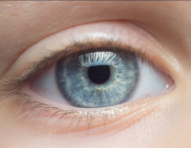
It is the main cause of dry eye, but diagnostic methods are limited.
MGD is the leading cause of evaporative dry eye, a condition that affects hundreds of millions of people worldwide. This disorder occurs when the lipid glands in the eyelids become blocked or malfunction, leading to an unstable tear film. Current diagnostic methods rely primarily on clinical observations and functional tests, which can be subjective and may not detect early-stage disease. Imaging tools such as meibography provide structural information but do not capture molecular or compositional changes in glandular tissue.
Capturing spectral “fingerprints” in milliseconds
To address this gap, researchers designed a small optical chip that can extract spectral features at the point of detection. This device integrates a high-density metasurface filter into a CMOS sensor. Each filter selectively modulates the incident light, allowing the system to perform optical calculations rather than digital post-processing during image acquisition. This architecture performs spectral measurements and feature extraction simultaneously, producing a complete spectral feature map within tens of milliseconds. In contrast, traditional hyperspectral imaging systems typically require mechanical scanning and acquisition times on the order of seconds.
Distinctive spectral features of diseased glands
The research team applied the system to pathological tissue sections with and without MGD and identified wavelength-dependent differences between the groups. In the visible range, MGD samples showed a higher spectral response, which is consistent with changes in hemoglobin-related optical properties that may reflect inflammation or changes in the microcirculation. In the near-infrared range, glandular regions showed signal changes related to lipid composition and tissue structure. These spectral features are also associated with clinical indicators of disease severity, suggesting potential relevance for objective assessment.
Improved accuracy over standard imaging
By using these spectral features for classification, the SCNN-based model achieved an average diagnostic accuracy of 96.22%. This was comparable to traditional hyperspectral imaging and significantly higher than models based on standard RGB images. Unlike RGB imaging, which only captures color and morphology, spectral approaches provide access to biochemical information, contributing to improved identification performance. In addition to accuracy, this chip also offers significant speed advantages, with acquisition times reduced by orders of magnitude compared to scan-based systems.
Toward real-time ophthalmological diagnosis
The chip is manufactured using a CMOS-compatible process and has a compact footprint, suggesting potential for integration into clinical imaging devices such as slit lamp microscopes. If this approach is further developed, it may be possible to quickly, objectively, and quantitatively assess meibomian gland function during routine eye examinations.
sauce:
China Photonics Society
Reference magazines:
DOI: 10.1186/s43074-026-00246-2


