image:
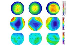
Although tomography plays a central role in measuring the overall corneal shape, its limitations create diagnostic gaps, especially in early detection. PS-OCT has the potential to address this issue by combining ultra-high resolution tomography with polarization-sensitive functional imaging. This focuses on the collagen tissue rather than just the thickness, helping to differentiate between true early disease and naturally thin but healthy corneas. Left column: Eyes classified as healthy. Mid-term, asymptomatic. Yes, keratoconus.
view more
Credit: RP Patil et al.
Keratoconus is a progressive eye disease that weakens and thins the cornea, the transparent front of the eye. In the early, asymptomatic stage, the cornea may still appear normal on routine examinations. However, at this time, especially when a patient is being evaluated for refractive surgery, an accurate diagnosis is paramount. A recent study found that combining polarization-sensitive optical coherence tomography (PS-OCT) with artificial intelligence reveals subtle corneal changes that are often missed by standard image processing. Published in Discovery of biophotonicsthis study is based on a large clinical dataset and points to new methods to improve early detection of keratoconus.
Why early detection is difficult
Most current screening tools rely on corneal geometry. Devices such as the Pentacam and MS-39 measure curvature, thickness, and surface roughness. These approaches work well once keratoconus is established, but are less reliable when disease-related changes are still microscopic. At this stage, the cornea may not yet show obvious deformation, even though the internal structure of the cornea has already changed. The authors therefore focused on a feature that shape-based imaging cannot directly capture: how collagen fibers inside the cornea are organized.
Measuring corneal structure using polarized light
PS-OCT is a high-resolution imaging technique that detects how polarized light changes as it passes through tissue. In the cornea, these changes reflect the alignment of collagen fibers, which is essential for mechanical strength. This breakdown of the collagen network is thought to occur early in keratoconus, before visible thinning or steepening occurs.
In this study, researchers used a custom-built PS‑OCT system that can resolve microscopic corneal layers. From each scan, they derived thickness maps of three layers: epithelium, Bowman’s layer, and stroma, as well as maps of phase lag, a measure related to collagen organization.
Comparison of three imaging approaches
The research team analyzed image data from 359 eyes examined at Narayana Nethralaya Eye Hospital. The dataset includes healthy eyes, eyes with clear keratoconus, and eyes classified as subclinical keratoconus based on established clinical criteria.
To fairly compare diagnostic performance, the authors trained three separate artificial intelligence models, each using data from one device: PS‑OCT, Pentacam, or MS‑39. All models used the same machine learning techniques and cross-validation strategy, allowing direct comparison of results.
How well did the model perform?
In healthy eyes and clear cases of keratoconus, all three models showed similar accuracy. Differences emerged in the asymptomatic group. Here, the PS‑OCT-based model classified more eyes as healthy compared to the two tomography-based models.
Importantly, these reclassifications were not random. Eyes identified as truly asymptomatic by PS‑OCT showed higher phase retardation values and subtle differences in subcorneal thickness, even when the overall corneal shape appeared normal. In contrast, eyes reclassified as healthy by PS-OCT tended to have collagen and layer thickness patterns that resembled normal corneas.
What the images reveal
A detailed case study highlights these differences. In healthy eyes, the phase retardation map was uniform and the thickness of Bowman’s layer was relatively uniform. Asymptomatic eyes had a gradual but consistent increase in central corneal phase delay with a slight thinning of Bowman’s layer. Eyes with established keratoconus showed a highly irregular pattern across all maps.
These findings suggest that PS-OCT is sensitive to microstructural differences that precede obvious shape changes. This technology provides a different perspective on corneal health by capturing both structural and polarization-based information.
Why is this important for patients?
A common clinical dilemma is how to manage patients with thin or slightly irregular but stable corneas. Current methods often label such eyes as “suspicious,” which can limit treatment options even when the risk of progression is low. This study shows that PS‑OCT can help distinguish true early disease from naturally thin but healthy corneas by focusing on collagen organization rather than thickness alone.
The authors stress that long-term studies are needed to confirm whether eyes reclassified as healthy remain stable over time. Still, their results demonstrate that polarization-sensitive imaging has the potential to improve reliability of early diagnosis and support safer decision-making in screening for refractive surgery.
Looking to the future
This study highlights the value of looking beyond the shape of the cornea by combining ultra-high resolution imaging and artificial intelligence. PS‑OCT does not replace existing tools, but complements them by adding information about tissue integrity that standard tomography cannot provide. As imaging technology continues to evolve, such approaches could help clinicians detect keratoconus earlier and tailor care more precisely to individual patients.
For more information, see the original Gold Open Access article “Advances in subclinical keratoconus detection using polarization-sensitive optical coherence tomography and artificial intelligence” by R. Patil et al. biophoton. discovery 3(1), 015004 (2026), doi 10.1117/1.BIOS.3.1.015004
journal
Discovery of biophotonics
Research theme
not applicable
Article title
Advances in the detection of asymptomatic keratoconus using polarization-sensitive optical coherence tomography and artificial intelligence
Article publication date
February 9, 2026
Disclaimer: AAAS and EurekAlert! We are not responsible for the accuracy of news releases posted on EurekAlert! Use of Information by Contributing Institutions or via the EurekAlert System.