There may be two different subtypes of multiple sclerosis, according to new research led by scientists at University College London (UCL). If the findings are validated, they could help doctors provide more specialized care to their patients.
The study used machine learning to analyze data from blood tests and brain scans of 634 patients who participated in two different clinical trials. Machine learning models are trained to detect subtle patterns that humans might miss.
The blood test was to detect a protein called serum neurofilament light chain (sNfL), a known biomarker for neurological diseases including multiple sclerosis (MS).
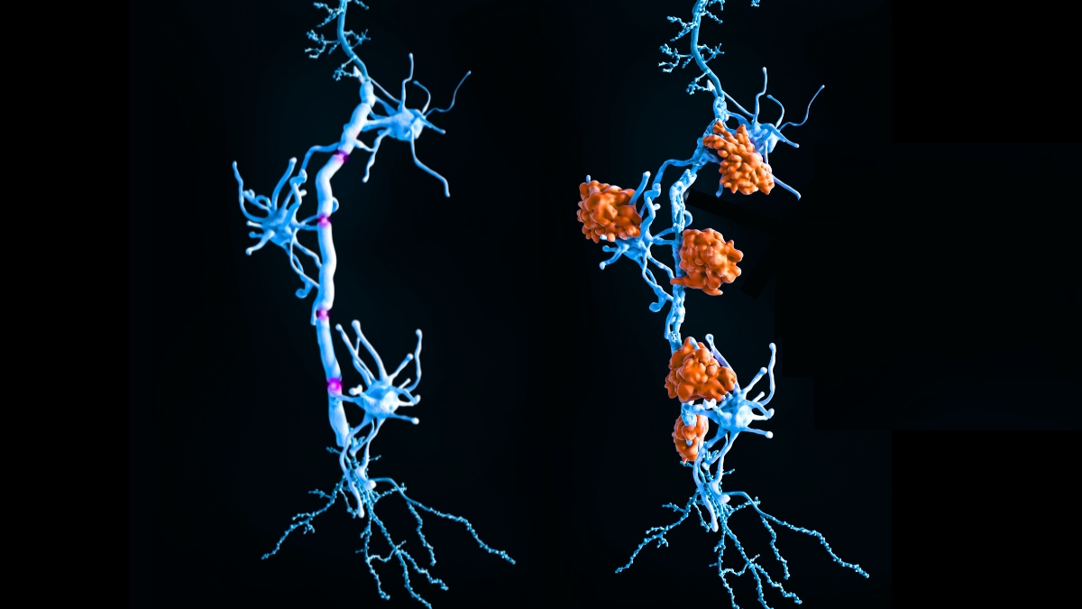
Meanwhile, MRI scans investigated damage and other changes in different parts of the brain. In MS, the body's immune system mistakenly attacks the protective sheath that surrounds nerve cells, leaving damage that prevents nerve transmission.
RELATED: Blood signals may predict multiple sclerosis seven years before symptoms
By comparing blood test results and brain scans, the model was able to classify patients into separate subtypes.
Patients classified as having “early sNfL” showed early elevated protein levels and damage to the corpus callosum, the structure that connects the left and right hemispheres of the brain. This subtype is more aggressive, and patients appear to develop brain lesions earlier than other subtypes.
The second subtype, termed “late-sNfL,” appeared to progress more slowly. The first sign in this group of patients was a shrinkage of the limbic cortex and gray matter deep in the brain. It wasn't until much later that sNfL levels in their serum began to rise.
is a neuroscientist at UCL and Queen Square Analytics is a research spin-off company.
“This helps clinicians understand where a person is in the disease pathway and who needs closer monitoring or early targeted treatment.”
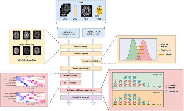
The machine learning model was trained on data from 189 patients with various types of MS (relapsing-remitting or secondary-progressive MS) and then tested on a further 445 patients recently diagnosed with the disease.
Neurofilaments are proteins that support neurons throughout the central and peripheral nervous systems, and in healthy patients, neurofilament turnover is considerably slower. However, due to neurodegeneration, these proteins are shed into body fluids at higher rates, making them potential biomarkers for diseases and disorders of the nervous system.

Unfortunately, in serum the difference is quite subtle, making it difficult to use for diagnosis. MRI scans can also reveal patterns in the spread of MS, but they cannot reveal the details of the disease.
The scientists behind the new study suggest that combining neurofilament levels with other data such as MRI scans could make their measurements more useful.
 frame border=”0″ permission=”accelerometer; autoplay; clipboard write; encrypted media; gyroscope; picture-in-picture. web-share” referrerpolicy=”strict-origin-when-cross-origin” allowedfullscreen>
“By adding sNfL, a well-established indicator of neuroaxonal damage, we have advanced beyond the structural snapshot provided by MRI alone,” the researchers conclude.
Currently, MS is classified and treated based on symptoms and disease progression, but the underlying mechanisms remain unexplained. Researchers in the new study say that if validated in future studies, this combination technology could help doctors recommend more appropriate treatments.
The study was published in the journal brain.


